Vol 8 No 3 (2013)
Original Article(s)
-
Background: Patients suffering from major beta thalassemia need frequent blood transfusions and, if not treated well, would be at risk of heart dysfunction. This study was performed to determine the diagnostic value of electrocardiography versus echocardiography in measuring the left ventricular mass index in these patients.
Methods: Between July 2010 and June 2011, 82 asymptomatic patients over 10 years of age with major thalassemia (42 men with a mean age of 17.65 ± 3.39 years and 40 women with a mean age of 16.9 ± 3.38 years) were enrolled in this study. For all the patients, standard electrocardiography (to measure R in aVL and S in V3 and calculate left ventricular mass index by electrocardiography) and echocardiography (to measure interventricular septum diameter in diastole, left ventricular posterior wall diameter in diastole, and left ventricular diameter in diastole in order to calculate left ventricular mass index by echocardiography) were performed, at least one week after transfusion. The calculated left ventricular mass indices were thereafter compared between the two methods (electrocardiography and echocardiography).
Results: Sensitivity, specificity, positive predictive value, and negative predictive value in the two techniques in determining the left ventricular mass index were 67%, 25%, 89%, and 7% in the females, 65%, 33%, 92%, and 6% in the males, and 67%, 14%, 89%, and 3% in the total population, respectively. Furthermore, this study demonstrated that the average left ventricular mass index by echocardiography and electrocardiography was 104.86 ± 21.65 gr/m2 and 91.69 ± 12.03 gr/m2, respectively. Echocardiography was much more accurate than electrocardiography in determining the left ventricular mass index (p value = 0.0001).
Conclusion: The findings of this study demonstrated that echocardiography was more accurate and more reliable than electrocardiography in determining the left ventricular mass index in major thalassemia patients. -
Background: During off-pump coronary artery bypass (OPCAB), the heart is subjected to ischemic and reperfusion injury. Preconditioning is a mechanism that permits the heart to tolerate myocardial ischemia. The aim of this study was to compare the effects of Adenosine preconditioning with ischemic preconditioning on the global ejection fraction (EF) in patients undergoing OPCAB.
Methods: In this single-blind, randomized controlled trial, sixty patients undergoing OPCAB were allocated into three equally-numbered groups through simple randomization: Adenosine group, ischemic group, and control group. The patients in the Adenosine group received an infusion of Adenosine. In the ischemic group, ischemic preconditioning was induced by the temporary occlusion of the left anterior descending coronary artery twice for a 2-minute period, followed by 3-minute reperfusion before bypass grafting of the first coronary vessel. The control group received an intravenous infusion of 0.9% saline. Blood samples at different times were sent for the measurement of creatine kinase isoenzyme MB (CK-MB) and cardiac troponin I (cTnI). We also recorded electrocardiographic indices and clinical parameters, including postoperative use of inotropic drugs and preoperative and postoperative EF.
Results: History of myocardial infarction, hyperlipidemia, diabetes mellitus, kidney disease, preoperative arrhythmias, and utilization of postoperative inotrope was the same between the three groups. The incidence of postoperative arrhythmias was not significant between the three groups. Also, there were no significant differences in preoperative and postoperative EF and the serum levels of enzymes (cTnI and CK-MB) between the groups.
Conclusion: Based on the findings of this study, there was no significant difference in the postoperative EF between the groups. Although the incidence of arrhythmias was higher in the ischemic preconditioning group than in the other groups, the difference between the groups did not constitute statistical significance. -
Background: Administration of glycoprotein IIb/IIIa inhibitors is an effective adjunctive treatment strategy during primary percutaneous coronary intervention (PPCI) for ST-segment elevation myocardial infarction (STEMI). Recent data suggest that an intracoronary administration of these drugs can increase the efficacy of PPCI. This study was done to find any potential difference in terms of efficacy of administering intracoronary Abciximab vs. intravenous Eptifibatide in primary PPCI.
Methods: A total of 40 STEMI patients who underwent PPCI within 12 hours of symptom onset were randomized to either an intracoronary Abciximab (0.25 µg/kg) bolus or two boluses of intravenous Eptifibatide (0.180 µg/kg) each 10 minutes. The primary end points were enzymatic infarct size, myocardial reperfusion measured as ST-segment resolution (STR), and post-procedural thrombolysis in myocardial infarction (TIMI) grade flow of the infarct-related artery. The secondary end points were intra-procedural adverse effect (arrhythmia) and no-reflow phenomenon, in-hospital mortality, reinfarction, hemorrhage, and post-procedural global systolic function.
Results: Post-procedural TIMI grade 3 flow was achieved in 95% and 90% of the intracoronary Abciximab and intravenous Eptifibatide groups, respectively (p value = 0.61). The infarct size, as assessed by the area under the curve of creatine phosphokinase-MB in the first 48 hours after PPCI (µmol/L/hr ), was similar between the intracoronary Abciximab and intravenous Eptifibatide groups: 6591 (interquartile range [IQR], 3006.0 to 11112.0) versus 7,294 (IQR, 3795.5 to 11803.5); p value = 0.59. Complete STR was achieved in 55% and 45% of the intracoronary Abciximab and intravenous Eptifibatide groups, respectively (p value = 0.87). No deaths, urgent revascularizations, reinfarctions, or TIMI major bleeding events were observed in either group.
Conclusion: The intracoronary administration of Abciximab was not superior to the intravenous administration of Eptifibatide in the STEMI patients who underwent primary PCI. -
Background: The existing evidence suggests that plasma adiponectin concentrations can be indicative of the presence and severity of coronary artery disease (CAD). However, the results of the studies conducted hitherto on this subject are inconsistent. We sought to investigate the possible correlation between plasma adiponectin levels and the presence and severity of CAD in patients undergoing non-urgent coronary angiography.
Methods: In 399 consecutive patients undergoing non-urgent coronary angiography for CAD survey, plasma adiponectin, triglyceride, total cholesterol, high-density lipoprotein and low-density lipoprotein cholesterol, and fasting blood sugar levels were measured and demographic characteristics such as age, sex, Body Mass Index, diabetes mellitus history, systemic hypertension history, and family history of CAD were collected. According to the angiography results, the patients were divided into two groups of CAD and non-CAD. The severity of coronary atherosclerosis in the CAD group was defined using the Gensini score system.
Results: Average age was 61.4 ± 9.94 years in the CAD group and 57.9 ± 10.75 years in the non-CAD group. Also, 73.7% of the CAD group and 55.4% of the non-CAD group were male. Totally, 278 (69.7%) patients were found to have CAD. Patients without CAD did not have higher mean plasma adiponectin concentrations than did those with CAD (13.38 ± 11.96 vs. 14.95 ± 14.11 mcg/ml; p value = 0. 896). After adjustment for CAD conventional risk factors, plasma adiponectin levels still were not associated with CAD. No association was found between plasma adiponectin levels and the Gensini score. Furthermore, in contrast to the fairly strong correlation previously reported, there was no correlation between adiponectin levels and conventional CAD risk factors.
Conclusion: We could not observe any relationship between plasma adiponectin concentrations and the presence or severity of CAD in patients undergoing coronary angiography. -
Background: Although percutaneous coronary intervention (PCI) improves outcomes compared to thrombolysis, a substantial number of ST-elevation myocardial infarction (STEMI) patients do not achieve optimal myocardial reperfusion. This study was designed to evaluate factors related to suboptimal myocardial reperfusion after primary PCI in patients with STEMI.
Methods: Totally, 155 patients (124 men; mean age = 56.6 ± 11.03 years, range = 31- 85 years) with STEMI undergoing primary PCI were retrospectively studied. Additionally, the relationships between the occurrence of reperfusion failure and variables such as age, sex, cardiac risk factors, family history, Body Mass Index, time of symptom onset, ejection fraction, previous PCI, coronary artery bypass graft surgery or previous myocardial infarction, and angiographic data were analyzed.
Results: Procedural success was 97.1% and complete ST resolution occurred in 43.2%. Age; cardiac risk factors; family history; body mass index; previous MI, coronary artery bypass graft surgery, or PCI; and use of thrombectomy device and GPIIb/IIIa inhibitor were not the determining factors (p value > 0.05). According to our multivariate analysis, time of symptom onset (OR [95% CI]: 045 [0.2 to 0.98]; p value = 0.044) and ejection fraction (OR [95% CI]:0.37 [0.26 to .091]; p value = 0.050) had reverse and male gender had direct significant associations with failed reperfusion (OR [95%CI]:0.34 [0.11 to 1.08]; p value = 0.068). More degrees of ST resolution occurred when the right coronary artery was the culpritvessel (p value = 0.001). The presence of more than three cardiac risk factors was associated with failed reperfusion (p value= 0.050).
Conclusion: Considering the initial risk profile of patients with acute STEMI, including time of symptom onset and ejection fraction, as well as the accumulation of cardiac risk factors in a given patient, we could predict failed myocardial reperfusion to design a more aggressive therapeutic strategy. -
Background: Serum N-terminal pro-brain natriuretic peptide (NT-proBNP), a polypeptide secreted by ventricular myocytes in response to stretch, was suggested as a predictor of adverse prognosis of the acute coronary syndrome (ACS). We examined the association between NT-proBNP level and angiographic findings in ACS patients to determine whether it could be used as a predictor of the severity of angiographic lesions.
Methods: This cross-sectional study was performed on 126 patients with chest pain or other ischemic heart symptoms suggestive of ACS. Venous blood samples were drawn to measure serum levels of NT-proBNP. Afterward, coronary angiography was performed and the patients were categorized into four groups according to the number of coronary vessels with significant stenosis. The severity of angiographic lesions was assessed with the Gensini scoring system.
Results: According to angiographic diagnosis, 11 (8.7%) patients had normal coronary arteries (no coronary artery disease [CAD]) and 115 (91.3%) had CAD, of whom 108 (85.7%) had obstructive CAD and 7 (5.6%) had minimal CAD. The serum NT-proBNP concentration was higher in the CAD group than in the non-CAD group (p value <0.01). A progressive significant increase in the NT-proBNP concentration according to the Gensini score and the number of involved vessels was reported after adjustment for sex and age. Furthermore, the Receiver Operating Characteristic Curve (ROC) analysis indicated that an NT-proBNP cut-point of 400 pg/ml could predict obstructive CAD with a sensitivity of 65% and a specificity of 78%.
Conclusion: Higher levels of NT-proBNP among our ACS patients were associated with the severity of angiographic lesions in terms of both the Gensini score and the number of involved vessels. This finding underscores the potential role of NT-proBNP in predicting the severity of CAD before performing angiography.
Case Report(s)
-
Bifid cardiac apex is a rare anomaly of human hearts. We report of the case of a 34-year-old man with a previous history of ventricular septal defect (VSD) and subvalvular pulmonary stenosis. He had undergone pulmonary commissurotomy and VSD closure 22 years before he was referred to our center for evaluation of progressive dyspnea. Transthoracic echocardiography revealed atrial septal defect (ASD), multiple VSDs, severe pulmonary regurgitation, and a bifid cardiac apex. The patient was referred for re-do surgery for ASD and VSD closure along with pulmonary valve replacement, but he refused the surgery.
-
Congenital anomalies of coronary arteries, albeit rare, may be significant contributors to angina pectoris, hemodynamic abnormalities, and sudden cardiac death. A 47-year-old man referred to us with atypical chest pain. Electrocardiography demonstrated no significant ischemic changes, but cardiac troponin I test was positive. The patient underwent coronary angiography, which revealed a single coronary artery from the left Valsalva sinus. In addition, the left anterior descending (LAD) and the left circumflex (LCx) arteries were in normal position with significant stenosis in the mid-portion of the LAD and the distal portion of the LCx. A large branch originated from the distal portion of the LCx and tapered toward the proximal portion as the right coronary artery (RCA). This is a rare coronary anomaly that has no ischemic result. Coronary lesions were the cause of the patient’s angina pectoris. Angioplasty and stenting of the LAD and LCx was done, and medical therapy (Clopidogrel, Aspirin, Atorvastatin, and Metoprolol) was continued. The patient was asymptomatic at 8 months’ follow-up.
-
Takotsubo cardiomyopathy (TCM), also known as stress-induced cardiomyopathy, is a clinical syndrome of transient left ventricular (LV) apical wall motion abnormality with relative preservation of the basal heart segments in the absence of any significant atherosclerosis. Recurrence of this condition is rare. We report a postmenopausal woman, who experienced two episodes of TCM within 4 months following emotional and physical stress. In the first episode, she was admitted due to severe dyspnea, accompanied by sudden-onset, prolonged, burning chest pain and palpitation. Transthoracic echocardiography revealed akinesia of the LV, with the exception of the basal regions. Coronary angiography demonstrated no significant coronary artery disease, and follow-up echocardiography showed normalization of the LV wall motion abnormalities. In the second episode, she experienced similar symptoms and echocardiography revealed similar changes. Multi-detector computed tomography revealed normal coronary arteries. After 9 days, she was discharged in good condition; and at 3 months’ follow- up, she was symptom-free with normal echocardiography.
Photo Clinic
-
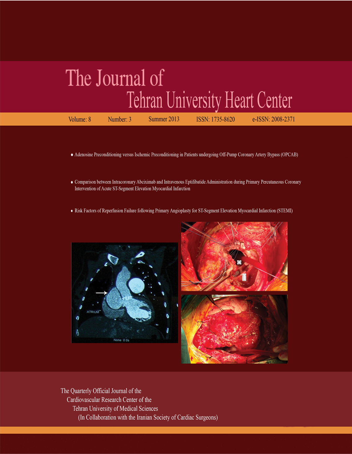
A 32-year-old female patient with previous Bentall operation and mitral valve repair surgery due to severe aortic insufficiency, mitral valve insufficiency, and ascending aortic aneurysm was admitted to our hospital with serious dyspnea, fatigue, and mild chest pain. Two-dimensional echocardiography demonstrated a markedly dilated basal aorta and cardiac chambers. Thoracic computed tomography scan highlighted a pseudoaneurysm, 14.5 cm in diameter (Figure 1). Urgent surgery was planned. The operation was performed under deep hypothermic cardiopulmonary bypass (arterial and venous line in the right femoral artery and vein). A large aortic pseudoaneurysm was demonstrated arising from the dehiscence of the proximal graft anastomosis (Figure 2). The composite graft did not require replacement, and it was possible to simply re-suture the composite graft and directly close the tear. The postoperative course was uneventful with no further evidence of leak from the anastomotic sites.


